
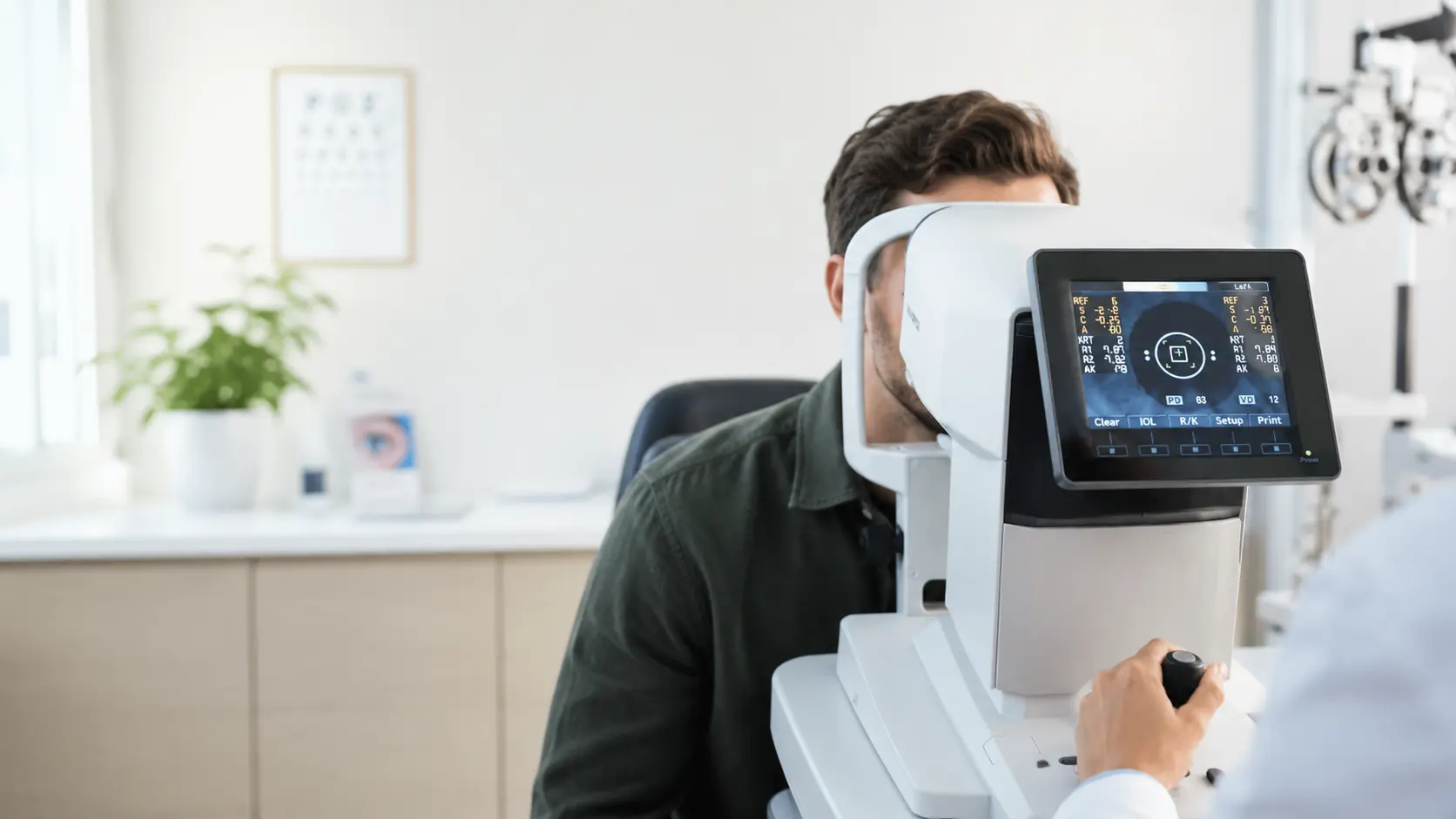
Advanced Diagnostic Technology in Pea Ridge
The best treatment starts with the best diagnosis. We've invested in modern diagnostic technology that catches eye problems years before symptoms appear — because early detection is the difference between managing a condition and losing vision to it.
What sets our advanced diagnostic technology apart.
The best treatment starts with the best diagnosis. We've invested in modern diagnostic technology that catches eye problems years before symptoms appear — because early detection is the difference between managing a condition and losing vision to it.
OCT (Optical Coherence Tomography)
A non-invasive scan that images the layers of your retina in microscopic detail — essential for glaucoma and AMD.
Wide-field retinal imaging
A panoramic photo of your retina that captures far more than a standard exam, documenting your eye health over time.
Visual field testing
Maps your full field of vision to detect blind spots and peripheral loss from glaucoma or neurological conditions.
Digital documentation
We image and store your results so we can compare year over year and catch subtle changes early.
Your advanced diagnostic technology visit.
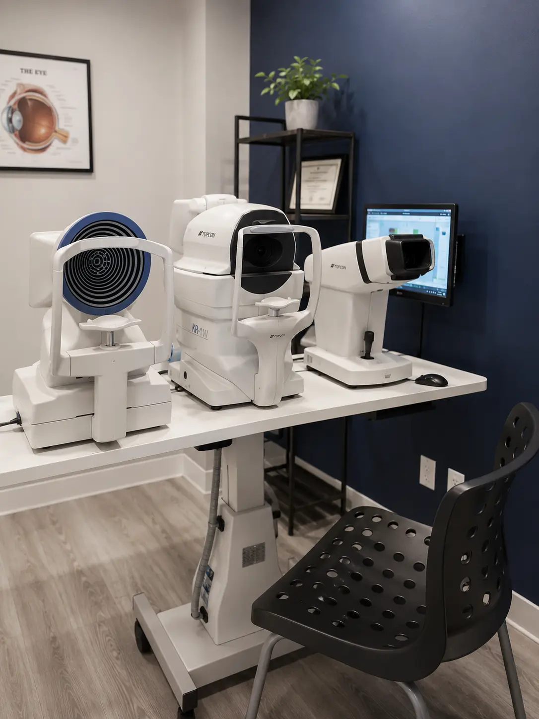
Diagnostic imaging is quick, painless, and often replaces the need for dilation. Scans take just minutes, and we review the results with you on-screen — so you can actually see what we see. These images become part of your record, letting us track your eye health over time.
Seeing problems years before symptoms.
The value of advanced imaging is simple: most serious eye diseases begin silently, and the earlier they are found, the more vision is preserved. OCT imaging works like an ultrasound made of light, capturing cross-sections of the retina and optic nerve in microscopic detail. It can reveal the earliest thinning of nerve tissue in glaucoma, subtle fluid or structural changes in macular degeneration, and diabetic changes in the retina, often years before any of them would be visible on a standard exam or noticeable to you.
Widefield retinal photography complements it by capturing a panoramic image of the retina in a single flash, documenting the far edges where tears, thinning, and other peripheral findings hide. Visual field testing maps your side vision with precision, which matters because diseases like glaucoma steal peripheral vision so gradually that the brain fills in the gaps.
The quiet superpower of all this technology is the baseline it creates. Eye disease is usually diagnosed from change over time rather than a single snapshot: an optic nerve that measures slightly thinner than it did two years ago, a retinal finding that has grown, pressure trending in the wrong direction. With years of scans on file at one practice, small changes stand out immediately and stable findings can be confidently left alone, which means fewer unnecessary specialist referrals and faster action when something real appears.
You will encounter this equipment as a routine part of a comprehensive eye exam when your eyes call for it, and it does the heavy lifting in our glaucoma, macular degeneration, and diabetic eye exam care. The scans are quick, painless, and often reduce the need for dilation, and Dr. Daiber reviews the images with you on screen so you can see what he sees.
Common questions about advanced diagnostic technology
Does OCT imaging replace dilation?+
In many cases, yes — OCT and wide-field retinal imaging can capture detailed views without dilation. We'll recommend dilation only when it adds diagnostic value.
Is retinal imaging covered by insurance?+
Coverage varies. When imaging is medically necessary (monitoring disease), it's typically covered by medical insurance. For routine screening, there may be a small fee — we'll always tell you first.
Why is early detection so important?+
Most serious eye diseases — glaucoma, AMD, diabetic retinopathy — cause no early symptoms. Imaging catches them before vision loss occurs, when treatment is most effective.
How often should I have diagnostic imaging?+
It depends on your age, risk factors, and any existing conditions. For most patients, baseline imaging plus periodic follow-up is ideal. We'll recommend a schedule.
Explore related services.
Ready to get started?
Book a advanced diagnostic technology appointment online or call us directly. New patients welcome.
Let's see what good vision can do.
Book online in under a minute, or call us directly. New patients welcome.